1. Introduction
Our eyes are constantly delivering a great deal of information to our visual
processing system. Although our visual field of view is fairly
large, we may not necessarily be aware of or focus on every location in
our field of view. On the contrary, we are only aware of and concentrate
on a small fraction of the available information. This process is
one of the functions of visual attention [10] and allows us to focus on
particular locations in our visual field thereby avoiding an overload of
the system. Such a selective process may appear to occur effortlessly,
immediate and we may not always be aware of it. However, there are
actually some complex processes occurring in the brain including the visual
cerebral cortex. The human and primate visual cerebral cortex can
be hierarchically divided into many distinct areas (see figures 1 and 2),
each representing some portion of the information in the visual scene [4].
These areas have been studies extensively throughout the year however,
our knowledge is still very incomplete. We do not know the exact
functions of each area, their interconnections, and whether there are actually
more areas we are not aware of yet. Recently, research is also taking
an interest as to how non-visual input, or "extraretinal sources"
(as referred to by the authors, for example, memory, motor planning,
attention), can affect the processing and the representations of these
areas. These studies have shown that neurons can be excited by extraretinal
sources and can therefore deliver non-visual signals. Plenty of psychophysical
studies related to attention have also been conducted and indicate that
attention has a "modulatory" influence (e.g. it can be switched,
it is selective [5], [1], [9] and have also discovered neural changes associated
with attention [2], [6]. Attention has been found to play a role
in form and color processing in areas V2 and V1 [7], [8], and has also
been detected in the dorsal and ventral pathways of the visual cortex [3].
1.1 Motivation of this studyDespite all the research related to attention,
few studies have focused on attention with respect to visual motion, despite
the fact that motion processing is an important part of vision! Furthermore,
understanding of the neural basis of motion analysis would be incomplete
without knowing how it's affected by the behavioral state [11]. Visual
motion processing in the visual cortex is primarily performed in areas
MT (Middle Temporal) and MST Medial Superior Temporal), both of which contain
many direction sensitive neurons.
The study described in this paper is concerned with examining the affect
of attention with regards to visual motion. It aims to examine the
importance of attentional modulation of visual motion signals, by examining
the response (using single cell recordings), of neurons in the areas MT
and MST of two macaque monkeys, under different attentional conditions.
So just what exactly does this hierarchy look like and where are the
areas MT and MST located? Figure 1 illustrates the flat map of the
macaque monkey as determined by Fellman and Van Essen [4].
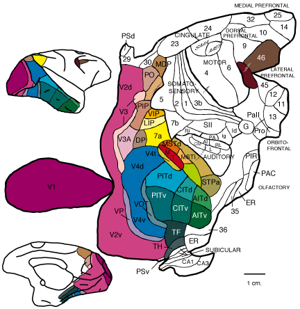 |
| Figure 1. Flat map of
the macaque monkey brain as determined by Fellman and Van Essen [4]. |
The hierarchy of areas, again as determined by Felleman and Van Essen
[4], is illustrated in figure 2.
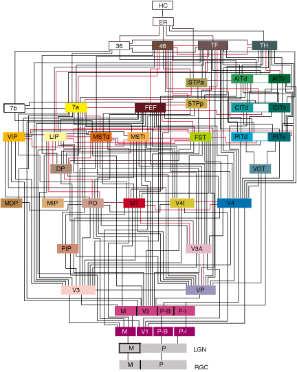 |
| Figure 2. Hierarchy
of the visual areas in the brain of a macaque monkey as determined by Fellman
and Van Essen [4]). |
Note; these diagrams were constructed over 10 years ago. Some
off the information may have been updated over time however, the work of
Felleman and Van Essen is still references in current papers including
this paper.

