3. Orienting - Visual Locations
Orienting to visual locations refers to moving our eyes and possibly head
such that some target (stimulus) falls on our fovea. It allows us
to attend to that location of the target. It is also possible to
attend to a location without actually moving our eyes or head (this is
known as covert attention whereas moving the eyes/head is referred to as
overt attention). Within the first 150ms of attending to some location,
we are able to respond faster to events occurring there and detect events
occurring at that location with lower thresholds. Actually, improvements
begin before any eye or head movements (shift of attention)
Furthermore monkey brain studies have shown increased neuronal responses
in the following areas, indicating their importance to attention.
-
Posterior parietal lobe
-
Lateral pulvinar nucleus
-
Superior colliculus
-
Parietal cortex
Further evidence of the role in attention of the above areas comes from
the study of primate and human brain lesion subjects who have suffered
damage to these areas. Lesions in any of these areas will reduce
the ability to shift attention to some other location. For example,
damage to the superior colliculus affects the ability to shift attention
to some other location regardless of whether we previously are or not attending
to some other target.
3.1 Hypothesis for the shifting of visual attention
Examining humans and animals with brain lesions, has confirmed that there
are definitely physical areas in the brain responsible for attention and
it has also provided an idea of the process required for the shifting of
visual attention. Based on these studies, the authors suggests the
following hypothesis for shifting of visual attention:
-
Parietal lobe disengages attention from the current focus.
-
Mid-brain area moves the "index of attention" to the target area.
-
Pulvinar collects data from the target location and processing begins.
Brain lesion studies have also revealed (although still controversial)
that that the left and right brain hemispheres have their own attentional
specialty. Both carry out the operations required to shift attention,
however, they differ with respect to the level of detail. "Large
and small" letter tests (e.g. forming large letters out of small
letters - see [6]) indicate that the right hemisphere is oriented towards
global processing whereas the left hemisphere is mainly responsible for
local processing. For example, the image of figure 3 illustrates
the combination of many small letter "L"s to form one large letter "L".
Global processing allows one to notice the single large "L" while local
processing will focus on the constituent small letter "L"s.
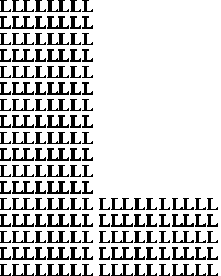 |
| Figure 3. Large letter
"L" constructed from many small letter "L"s. |
3.2 Pattern recognition - Visual Search
Attention is used in pattern recognition tasks from detecting features
to storing new visual patterns. An example of a pattern recognition
task is shown in figure x, where the task may be to locate the single red
triangle in the presence of other different color shapes.
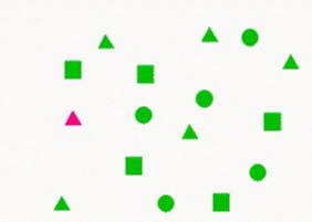 |
| Figure 4. Pattern recognition
task involving a single target shape with one distinct feature. Reprinted
from [10] |
In these pattern recognition tasks, increasing the number of non-targets
and their features does not influence the search for a one feature (e.g.
color, or shape as in figure 4 where the red target is separated by all
other non-target shapes by color only). However, increasing the number
of target features to more than one, does decrease the speed of the task.
For example, consider the example shown in figure 5. Finding the
red triangle (target) now takes more time than finding the same target
triangle from the example of figure 4..
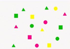 |
| Figure 5. Pattern recognition
task involving a multiple feature target. Reprinted from [10]. |
Further evidence that attention is involved in pattern recognition comes
from studies involving single cell recordings. Such studies indicate
the attention system interacts with the pattern recognition system through
the thalamus, requiring approximately 90ms to do so.


